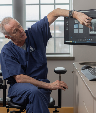
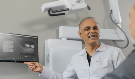
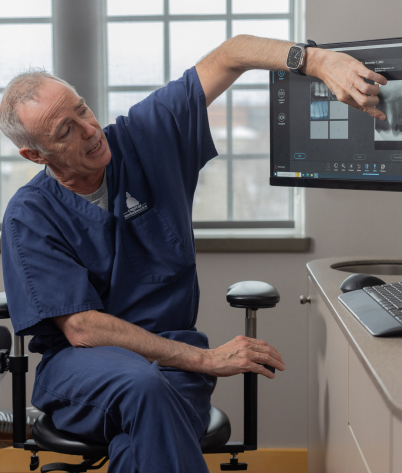
Introducing the Future of Intraoral Imaging
More information will lead to better clinical decisions
Dentistry has seen many innovations in recent history, but dental imaging has remained relatively unchanged for over 70 years. The PORTRAY System uses Tomosynthesis to produce dynamic 3D images that can be scrolled through in “slices” to show the inside of the teeth or other dental anatomy. This game-changing process provides dental practitioners with more data than ever before, allowing them to provide a new level of patient care.
The PORTRAY System takes multiple images from several different angles. The software then compiles those images into a 3D volume and dissects the volume into .5mm slices. This allows the dentist to scroll through the slices with a mouse to diagnose problems that are otherwise obscured or not clearly visible in a conventional 2D X-ray.
Conventional 2D X-rays
For the last century, dentists have relied on conventional 2D intraoral dental imaging systems. Dentists are still working with this technology, compelling them to rely on incomplete information. In many cases, dental conditions have required a wait-and-see approach until a prognosis was more evident, which could lead to a more invasive procedure.
The PORTRAY® System 3D Intraoral Tomosynthesis
The PORTRAY System gives dentists the power to see the unseen, obtaining more image data and viewing angles than ever before possible with intraoral imaging. With scrollable 3D images that let you view sections of the teeth in slices, the PORTRAY System helps dentists identify issues earlier. In addition to 3D imaging, the PORTRAY System offers a synthetic 2D mode for visibility into interproximal contacts.
The PORTRAY System is the first intraoral dental imaging device that can help clinicians to observe palatal roots and full canals that may have been previously invisible in a conventional 2D X-ray.
The PORTRAY System‘s rectangular cone magnetically registers with the sensor holder allowing alignment with the intraoral sensor, so the software knows where the sensor is in space.
Industry-Changing Software
Conventional 2D, 3D Tomosynthesis, and Synthetic 2D modes
Identify caries, cracks, and other pathologies that were previously inconclusive or invisible. Use 3D Tomo mode to bring obscure details into focus by scrolling through data-rich image slices. Use Synthetic 2D mode to rotate images to open contacts that appear closed on flat conventional 2D images.
3D Tomo takes multiple images from different angles. The software then compiles those images into a 3D volume and dissects that volume into .5mm slices. This allows dentists to scroll through the slices with a mouse to diagnose problems that are otherwise obscured or not clearly visible in a conventional 2D X-ray.
The Right Tools for the Job
The PORTRAY™ Software delivers new features without a difficult learning curve.
Adjust contrast, zoom in, and take measurements on images with the intuitive set of tools like the loupe tool, the contrast inversion tool, and the ability to compare older images to the 3D Tomo images side-by-side.
Connect
The PORTRAY Software integrates with the most popular patient management systems. It can also be used in stand-alone mode.
Industry-Changing Software
Conventional 2D, 3D Tomosynthesis, and Synthetic 2D modes
Identify caries, cracks, and other pathologies that were previously inconclusive or invisible. Use 3D Tomo mode to bring obscure details into focus by scrolling through data-rich image slices. Use Synthetic 2D mode to rotate images to open contacts that appear closed on flat conventional 2D images.
3D Tomo takes multiple images from different angles. The software compiles those images into a 3D volume, dissects that volume into .5mm slices. This allows dentists to scroll through the slices with a mouse to diagnose problems that are otherwise obscured or not clearly visible in a conventional 2D X-ray.
The Right Tools for the Job
The PORTRAY™ Software delivers new features without a difficult learning curve.
Adjust contrast, zoom in, and take measurements on images with the intuitive set of tools like the loupe tool, the contrast inversion tool, and the ability to compare older images to the 3D Tomo images side-by-side.
Connect
The PORTRAY Software integrates with the most popular patient management systems. It can also be used in stand-alone mode.
Better Information Leads to Better Patient Acceptance
The PORTRAY® System offers dentists the first opportunity to bring 3D Tomo to their practices, dramatically increasing the information they can gather during an exam. With the power of 3D Tomo, dentists can identify more pathology resulting in more actionable diagnoses. The imaging power of the PORTRAY System can be used to demonstrate the importance of procedures to patients. Payors will welcome the improved ability to confirm the dentist’s decisions.
Integrating the PORTRAY System into a practice is simple. The design maintains a similar workflow and fits into a similar space to that of conventional intraoral devices.
“In 70 years, this is the first viable X-ray device that changes how clinicians see dental structures on intraoral images.”
DR. ENRIQUE PLATIN
Retired professor of Oral and Maxillofacial Radiology, UNC at Chapel Hill, Adams School of Dentistry
“In 70 years, this is the first viable X-ray device that changes how clinicians see dental structures on intraoral images.”
DR. ENRIQUE PLATIN
Retired professor of Oral and Maxillofacial Radiology, UNC at Chapel Hill, Adams School of Dentistry